


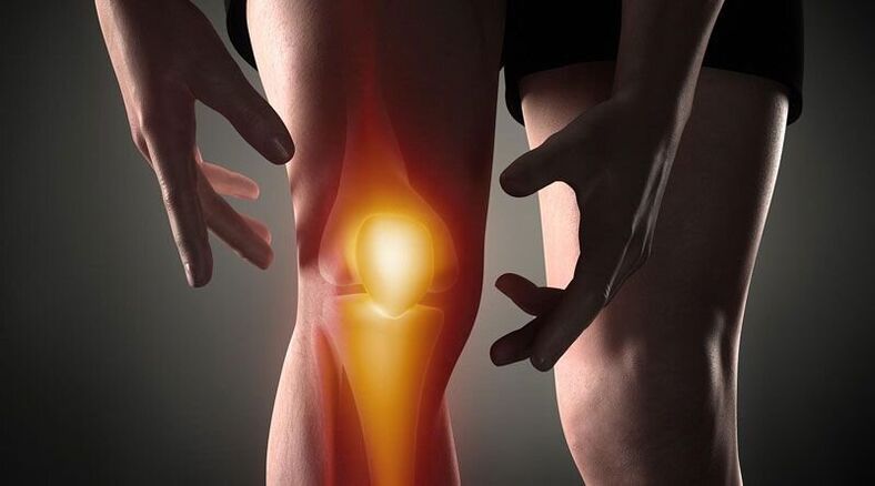
Knee pain-This is a sign of a pathological process affecting the cartilage, bone or soft tissue structure of the femoral-tibial and femoral-patella joints. Arthralgia can be based on trauma, inflammatory and degenerative diseases of the articular apparatus and periarticular structure. Patients may complain of sharp pain, soreness, burning, throbbing and other types of pain that occur during rest or when moving, supporting, bending and extending the leg at the knee. Diagnostics of causal pathology include instrumental imaging methods (Rg, ultrasound, CT or MRI, arthroscopy), joint capsule puncture, biochemical and immunological analysis. Until the diagnosis is clarified, rest, joint immobilization, NSAIDs and analgesics are recommended.
Causes of knee pain
Traumatic injuries
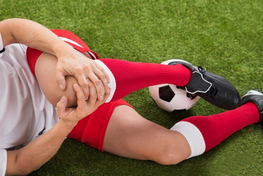
Usually they are the result of domestic trauma, often found in athletes: runners, jumpers, participants in playing sports. Developed by a fall, direct impact or twisting of the foot. Manifested by sharp pain at the time of injury. In the future, the pain syndrome becomes less pronounced, accompanied by an increase in edema. Abrasions and bruises are possible. As the frequency increased, the following injuries were identified:
- Knee injury. . . Occurs when falling to the knee or being hit directly. At first, the pain is sharp, hot, sometimes burning, but bearable, later - dull, aching, aggravated by movement. Bruising is possible. Foot support is preserved. Sometimes knee injuries are complicated by hemarthrosis, in such cases, the joint gradually increases in volume, becomes spherical, a feeling of pressure or rupture is added to the pain syndrome.
- Ligament rupture.It is found after twisting the leg, twisting forcibly, bending or overly extending in a non -physiological position. The painful sensation is stronger than with bruising; simultaneously with the onset of pain, one can feel how something is torn (much like how normal tissue is torn). It is accompanied by significant limitation of movement, support, twisting of the limbs, rapidly increasing hemarthrosis.
- Intra-articular fracture. . . They are detected by bumps, falls and twisting of the legs. In the case of an injury, a person feels a very sharp pain, often unbearable, sometimes a buzzing sound is heard. Patients themselves with intra-articular fractures describe their feelings as follows: "the pain is such that it is dark in the eyes, the world no longer exists, you do not understand anything. " After that, the pain becomes less severe, but remains of high intensity. Support is usually impossible, movement is almost completely limited. Edema and hemarthrosis develop rapidly.
- Dislocation.Is it the result of a blow or a fall to the knee. At the time of dislocation of the patella, a sharp pain occurs, accompanied by a feeling of bending of the foot and displacement in the knee. No movement is possible, reference function can be saved. On the anterior surface of the knee, a clear deformation is visible, which is then slippery due to the increasing edema. Sometimes hemarthrosis joins.
- Pathological fractures. They develop with minor injuries, are the result of a decrease in bone strength in osteoporosis, osteomyelitis, tuberculosis, bone tumors. The pain is aching, dull, reminiscent of pain syndrome with bruising. Signs that indicate a pathological fracture are the limitation or impossibility of support in the leg, a feeling of instability in the knee, sometimes deformity, dryness of the bones during movement.
- Damage to the meniscus.Tears of the meniscus are formed during twisting, shock, strong forced bending or knee extension, sharp turns with the foot fixed. At first, one feels a special click and a sharp shot pain in the depths of the joint. Then the pain somewhat diminishes, but becomes diffuse, sometimes - burning, breaking, intensifying when trying to support and move. Knee volume increases due to edema and hemarthrosis. Support becomes impossible, movement is very limited.
Inflammatory pathology
They can be contagious and non -contagious (post -traumatic, toxic allergies, metabolic, after vaccination). An abundant blood supply to the synovial membrane and periarticular tissues promotes the rapid development of inflammation in response to direct and indirect effects, and a large number of nerve endings cause significant pain reactions. The inflammatory process is often accompanied by synovitis (accumulation of aseptic fluid in the joints), with infection, pus may accumulate.
- Joint pain.Gonarthritis occurs after injury, sometimes complicating an infectious disease, detected in rheumatic diseases. May be acute or chronic. Knee pain is usually dull, aching, pressing or pulling. At first, the pain is not intense and intermittent, increasing in the evening or after exercise. Then the pain begins to merge, the intensity and duration of the pain syndrome increases. The joints swell, the skin on them turns red, the temperature rises. With synovitis, the contour of the knee is smoothed, there is a feeling of rupture. With suppuration, the severity of pain increases sharply, they become wrinkled, preventing sleep.
- Synovitis.It is not disease independent, complicating many acute and chronic pathologies of the joints. It forms in a matter of hours or days. At first, the pain is insignificant or absent, a feeling of fullness occurs. The knee is spherical, with a large amount of fluid, shiny skin. Movement is quite limited. When infected, the pain becomes noticeable, throbbing, twitching, intensifying with little movement and touch.
- Bursitis.Inflammation of the joint capsule located in the patella and popliteal fossa usually occurs when the knee is overloaded and the injury is repeated (e. g. , with constant support on the knee). With bursitis, the pain is local, dull, not intense, appears at a certain position of the limb, after a characteristic load, decreases when the position of the foot changes, massaging the affected area. If the posterior bag is affected, a painful sensation may occur while ascending or descending stairs. Minor local edema is sometimes determined. With bursa pus, the pain becomes sharp, wrinkled, burning, combined with hyperemia, edema of the affected area, symptoms of general intoxication.
- Tendinitis.Usually it is detected in overweight men and athletes, it affects the patella ligament itself. Initially, pain syndrome appears only with very intense effort, then with standard sports loads, then with daily physical activity or rest. Pain with tendinitis localized in front just below the knee, dull, exciting, with disease progression, sometimes paroxysmal, in some cases accompanied by mild redness and swelling, aggravated by pressure. Movement is usually full, less often slightly limited. Torn or ruptured ligaments may be caused by a decrease in their strength.
- Lipoarthritis.Hoff’s disease affects the layer of adipose tissue located below the patella. It is observed with persistent knee overload or as a result of long -term injuries. More often it affects athletes, older women. Someone complains of dull aching pain in combination, some advanced limits. With the severity of the pathology, the pain begins to interfere at night, there is a feeling of instability of the knee, subdued legs. When pressing on the side of the patella, a cracking sound or a soft creak is heard.
Autoimmune processes
The cause of diseases of this group is the production of antibodies to normal cells of the body with the development of aseptic inflammation of the immunocomplex of synovial membranes and cartilage, the phenomenon of vasculitis. Pathologies in most cases are chronic, without treatment they are prone to progression, and are often the cause of disability.
- Rheumatoid arthritis.Defeat is usually two -way. With minimal activity of autoimmune processes, weak or moderate pain, intermittent, pulling, pressing, accompanied by morning stiffness. With moderate activity, patients complain of prolonged periodic pain, pressing or bursting with moderate intensity, not only during movement, but also during rest. There is stiffness for hours, mild recurrent synovitis. With high activity of rheumatoid arthritis, the pain is strong, diffuse, exhausting, wavy in nature, increasing in the early hours of the morning. Stiffness becomes constant, a large amount of fluid accumulates in the knee, contractures form over time.
- Systemic lupus erythematosus.Arthralgia is often symmetrical, although one joint may be affected. It can occur at any stage of the disease; with recurrent courses of SLE, they resemble rheumatoid arthritis. With low process activity, the pain is short -term, not intense, local, painful, exciting. In severe cases, the pain syndrome develops, the pain is wavy, interferes with night sleep, becomes prolonged, diffuse, increases with movement, combined with synovitis, edema, hyperemia.
- Joint pain.Joint pain is one of the first manifestations of rheumatic fever, appearing 5-15 days after an acute infection, affecting several joints at once (usually in pairs). The pain is rather short -lived, but intense, migrating from one joint to another, differing in nature from pulling or pressing to burning or throbbing. The knees are swollen, hot, the skin on them is red. Movement is very limited. After a few days, the severity of the pain decreased, movement was restored. In some patients, residual effects in the form of moderate or mild dull pain persist for a long time.
- Reactive arthritis.More often occurs 2-4 weeks after intestinal and urogenital infections, usually affecting one or two joints of the lower legs, combined with urethritis, conjunctivitis. The development of reactive arthritis is preceded by increased urination, pain and a burning sensation in the urethra, lacrimation, and cramping in the eyes. Pain in the knee is strong or moderate, persistent, wavy, aching, pulling, wrinkling, combined with limitation of movement, deterioration of general condition, fever, severe swelling and redness of the affected area. The painful sensation and signs of inflammation persist from 3 months to 1 year, and then gradually disappear.
Degenerative-dystrophic process
They develop as a result of metabolic disorders in the soft tissue structures of the joints and periarticular. They have a chronic course, developing over the years. Often accompanied by the formation of calcifications, cysts and osteophytes, deformation of the knee surface. With significant destruction of the articular surface, it leads to significant deterioration of movement and support function, becomes a cause of disability, and requires the installation of an endoprosthesis.
- Osteoarthritis.It develops for no apparent reason or against a background of various injuries and diseases, especially in older and middle -aged people. Initially, the pain is mild, short -term, usually exciting or painful, occurs with prolonged effort and disappears during rest, often accompanied by cramping. Gradually the pain syndrome increased, the knees began to ache "in the weather" and at night, there was limitation of movement. Distinctive features of gonarthrosis are pain onset (pain until you "disperse"), periodic attacks of sharp cuts, burning pain or shooting as a result of blockage. During periods of exacerbation, synovitis often occurs, in which the pain becomes constant, pressing, ruptured.
- Meniscopathy. . . Usually detected in athletes, people whose work involves a significant load on the knee joint. Manifested by localized internal pain in the knee at the level of the joint space, more often on the outside of the knee. The pain increases during movement and subsides during rest, it can be dull, pressing or pulling. With progression, there is acute shooting pain when trying to move. On the anterolateral surface of the joint in the pain projection, a small painful formation is sometimes felt.
- Tendopathy. . . Tendons near the knee are affected. In the early stages, they are indicated by short -term local superficial pain at the peak of physical activity. After that, a painful sensation arises moderately, and then a light load, limiting normal daily activities. Exciting or aching pain, directly related to active movement, is not detected during passive extension and flexion of the knee, sometimes accompanied by dryness or tingling. In the lesion area, it is possible to investigate the site of the most severe pain. Signs of local inflammation (edema, hyperemia, hyperthermia) are insignificant or absent.
- Osteochondropathy.Children and young people are more often affected, the duration of the disease is several years. Usually they begin gradually with mild or intermittent lameness, dull pain that is not intense, aggravated by energy, disappearing during rest. With the development of osteochondropathy, the pain becomes intense, persistent, pressing, burning or burning, accompanied by severe lameness, limitation of movement and difficulty in resting the limbs. Then the pain gradually subsides, support function is restored.
- Chondromatosis.Usually diagnosed in older men, less frequently in infants. Joint chondromatosis is manifested by pain like a simple dull wave, often worsening at night and in the morning. Movement is limited, accompanied by cramps. Occasionally obstruction occurs, characterized by sudden sharp shooting pain, impossibility or severe limitation of movement. With the development of synovitis, the pain acquires a ruptured character, combined with an increase in knee volume, soft tissue swelling, and an increase in local temperature.
Tumors and tumor -like formations
Pain syndrome can be caused by cysts, benign or malignant tumors that directly affect joint or periarticular tissue. In addition, knee pain can serve as a worrying signal of hypertrophic arthropathy, paracancrotic polyarthritis - a characteristic paraneoplastic syndrome of lung cancer, breast cancer and other oncological processes.
- Cyst Baker.Represents a hernia protrusion in the popliteal fossa. In the early stages, it manifests itself as an unpleasant sensation or mild local pain along the back of the knee. Against the background of an increase in Baker cysts due to compression of nearby nerves, burning or shooting pain, numbness or tingling in a single area may appear. The symptoms are worse when trying to bend the knee as much as possible. In the popliteal fossa, an elastic and painful tumor-like formation is sometimes felt.
- Benign tumor.Includes chondromas, osteochondromas, non -ophthalmic fibromas and other neoplasms. They are characterized by a prolonged asymptomatic or low-symptom course, they may manifest themselves with vague and intermittent local pain. With large neoplasia, solid formation is felt, sometimes synovitis develops.
- Malignant neoplasia.The most common malignant tumors that affect the joint area are synovial sarcoma, osteosarcoma, and chondrosarcoma. They manifest with a dull local vague pain, sometimes with a certain circadian rhythm (worse at night). The intensity of pain increases, they become sharp, cutting, burning or wrinkled, spread along the knee and adjacent tissues, accompanied by deformation, edema, synovitis, expansion of the saphenous vein, violation of the general condition, the formation of contractures. Upon palpation, a painful tumor -like formation is determined. When the process begins, the pain is very painful, unbearable, exhausting, prevents you from sleeping, and is not eliminated by non -narcotic analgesics.
Invasive surgery and manipulation
The pain syndrome is triggered by damage to the knee tissue during an invasive procedure. The severity of pain directly depends on the manipulative trauma of the knee joint. With the penetration of pathogenic microbes into the joint area, pain is caused by inflammatory changes.
- Manipulation.The most common procedure is puncture. Post -puncture pain is short -term, not intense, rapidly subsiding, localized in the projection of the puncture, usually performed on the outer surface of the knee. After the biopsy, the pain may wrinkle at first, then become dull and disappear after a few days.
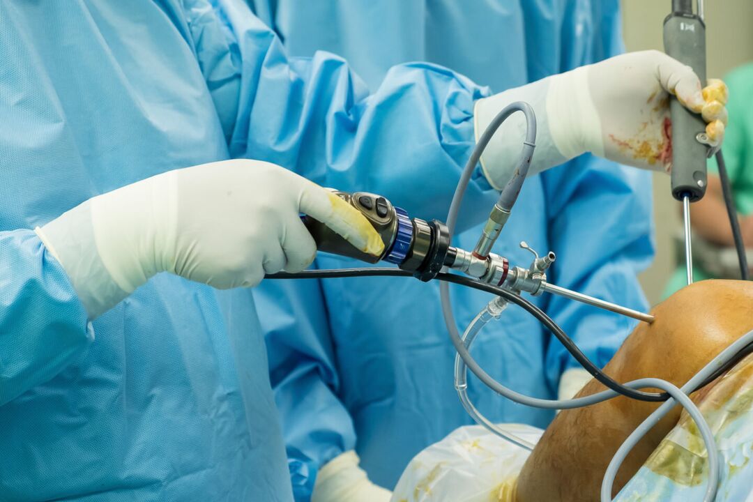
- Operation.After arthroscopy, the pain is mild, initially quite acute, then dull, subsiding after a few days or 1-2 weeks. After arthrotomy, the pain syndrome is more intense, it can persist for up to several weeks due to significant tissue damage. Usually, in the first 2 or 3 days after the intervention, the patient is given analgesics, then the pain becomes weak and gradually disappears.
Psychosomatic conditions
Sometimes arthralgia in the knee occurs in the absence of organic basis (trauma, inflammation, destruction, etc. ) under the influence of psychological factors. It is believed that such pain plays a protective role, as it helps reduce emotional stress by turning the experience into a physical sensation. The hallmarks of such pain are its uncertain nature, inconsistency, absence of visible changes, a clear relationship with physical activity and other factors that provoke the objective. Meteoropathic arthralgia is observed in people who are sensitive to changes in atmospheric pressure.
In addition, radiating pain in the knee is possible with coxarthrosis, lumbar osteochondrosis, Perthes disease, fibromyalgia, sciatic nerve neuropathy. However, with this pathology, other localization pain syndromes usually come to the fore. Additional risk factors that increase the likelihood of knee joint injury and disease include overweight, professional sports, hypovitaminosis, metabolic disorders, and old age. Hypothermia, stress, physical exercise, and dietary disorders can be provoking factors for the exacerbation of chronic pain.
Review
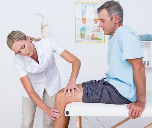
The diagnostic search algorithm is based on taking into account the nature of the pain syndrome, its duration, identifying symptoms and corresponding events before the onset of knee pain. At the first visit to the doctor (traumatologist-orthopedic, surgeon, rheumatologist), visual examination and palpation of the knee, assessment of the amount of active and passive movements is performed. Taking into account the data obtained, in the future, patients can be assigned:
- Laboratory blood tests. . . A complete blood count helps to identify hematological changes characteristic of infectious processes and acute inflammation (leukocytosis, increased ESR), eosinophilia, typical of allergic reactions. Biochemical and serological studies are the most informative for autoimmune diseases, characterized by the formation of certain acute phase proteins and immunoglobulins (CRP, rheumatoid factor, ASL-O, CEC, antibodies to DNA, etc. ).
- Radiography.The basic diagnostic method is X-ray of the knee joint in 2 projections. The presence of pathology is indicated by changes in the contour of the articular head and cavity, narrowing of the joint space, changes in the thickness of the end plate, the presence of edge defects at the articular ends of the bones, osteolysis and bone destruction. . In some diseases (meniscus trauma, Baker cyst), contrast arthrography shows the greatest sensitivity.
- Arthrosonography. . . Knee ultrasound is a fast, inexpensive, affordable and highly informative diagnostic method. Allows you to assess the presence of effusions and free bodies in the joint cavity, to identify damage and pathological changes in the periarticular soft tissues (signs of calcification, bleeding, etc. ). They help to distinguish with high accuracy the etiology of joint pain.
- CT and MRI. . . They are the method of choice for arthropathy of any genesis. They are used for a more detailed assessment of the nature and extent of pathological changes, to identify typical signs of traumatic lesions, inflammation and tumors of bone and soft tissue structures. CT and MRI of the joints are commonly used with the limited information content of other instrumental studies.
- Joint puncture. . . It is performed when there are signs of exudate or transudate accumulation in the joint capsule. As part of the differential diagnosis of inflammatory, degenerative and tumor diseases, cytological, bacteriological or immunological studies of synovial fluid are performed. To establish the diagnosis of autoimmune damage to the knee joint, tuberculous arthritis, synovioma, it is very important to carry out a biopsy of the synovial membrane.
- Arthroscopy. . . The purpose of invasive endoscopic diagnostics can be biopsy sampling, clarification of diagnostic information required during visual examination of joint elements. In some cases, diagnostic arthroscopy develops into therapy (removal of intra-articular body atroscopy, menissectomy, ligament autoplasty, etc. ).
Symptomatic treatment
Treatment of the cause of knee pain is carried out differently, taking into account the identified disease. At the same time, symptomatic care is an important part of a comprehensive treatment process aimed at reducing discomfort and improving quality of life. Immediately after the injury, it is recommended to apply cold compresses to the knee area - this will help reduce pain sensitivity. Ethyl chloride has a local cooling and anesthetic effect. In all cases, knee rest helps reduce pain. It is necessary to limit movement, give the legs a position where pain is minimal. When walking, a fixation bandage is applied to the knee, immobilization of the limb is possible with the help of a plaster cast.
In the acute period of injury or illness, it is strictly forbidden to massage the knee, use heating compresses, and wear high -heeled shoes. The main classes of drugs used for the treatment of pain and inflammatory symptoms are analgesics and NSAIDs in the form of ointments, tablets and injections. The measures listed can only temporarily reduce pain, but do not eliminate the cause of arthralgia. Therefore, all cases of knee pain require qualified diagnosis and treatment, and some conditions (fractures, dislocations, hemarthrosis) require emergency medical treatment. You can not delay a visit to the doctor if the pain is combined with changes in the shape of the knee (swelling, smoothing of contours, asymmetry), inability to perform flexion-extensor movements, patella votes, impaired limb support.